
Begin with the imaging principle
FDA explains that diagnostic ultrasound forms images using high-frequency sound waves and their echoes. It differs from imaging that uses ionising radiation. That difference does not make every use unlimited or risk-free: FDA discusses prudent use by appropriately trained healthcare providers. An old claim that a wireless probe needs no operator is not a description of responsible clinical assessment.
Draw the separate connections
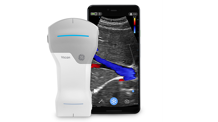
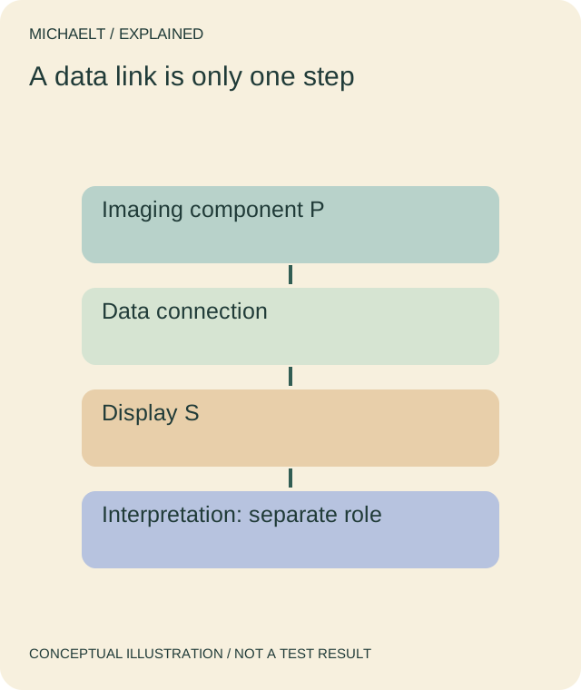
A conceptual diagram can show an imaging component, a data connection and a display. Wireless describes a connection; it does not explain whether an examination is clinically indicated, how the image was obtained or who interprets it. Do not infer autonomous diagnosis from the absence of a cable. The actual product’s intended use and instructions define its scope, not a stock photograph of a phone.
Keep the report and operator in the record
For a documentation enquiry, ask the responsible service to identify the actual device and professional process relevant to the examination. Keep image acquisition, a displayed image, a report and clinical interpretation as separate records. A connectivity demonstration does not validate an image or establish a diagnosis. This page provides no scanning, positioning, settings or image-reading instructions.
Retain the intended-use boundary
Ask an appropriately qualified professional about any individual imaging question, including pregnancy or an unclear indication. FDA discourages ultrasound used solely for non-medical fetal keepsake purposes. Do not use a general wireless-device article to perform a scan or declare a particular examination necessary or safe. The diagram explains relationships, not a complete clinical workflow or an approved product architecture.
Read the drawing as text
A conceptual connection from an imaging component to a screen does not establish a clinical interpretation or autonomous diagnosis.
Work through an example
A fictional listing shows probe P sending an image to screen S and calls it automatic diagnosis. Its supplied document establishes only a data connection; the professional interpretation process is not identified. The reader marks that missing link instead of treating the image as a diagnosis. No scan, patient, device approval, clinical result or artificial-intelligence capability is demonstrated.
Illustrative example; figures, people and organisations in the worked record are fictional.
The example record
Imaging
- Illustrative evidence
- Sound-based process
- Meaning or next step
- Not an operating tutorial
Connection
- Illustrative evidence
- P → screen S
- Meaning or next step
- Wireless is a transport property
Interpretation
- Illustrative evidence
- Professional process unidentified
- Meaning or next step
- Image alone is not diagnosis
Scope
- Illustrative evidence
- Actual intended use requested
- Meaning or next step
- No scan or approval established
Download blank worksheet (.csv) ↗Download worked example (.csv) ↗
Keep your own record
Use the result within its scope
Use the actual clinical service, device instructions and qualified advice. A connection diagram cannot authorise an examination, validate diagnostic performance or replace a trained healthcare professional.